Image: Difference between revisions
No edit summary |
No edit summary |
||
| Line 14: | Line 14: | ||
The value of each image pixel is in relative units. | The value of each image pixel is in relative units. | ||
== Color == | |||
It is important to note that '''<u>most</u>''' microscope | |||
Typically, microscope images are rendered in a false color | |||
Latest revision as of 19:28, 12 November 2024
An image is a visual representation of the biological system under study. Typically, these images are digital and viewed on a computer.
How microscope images are formed

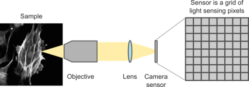
Images are formed by focusing light from the sample on to a detector, such as a camera. For this section, we will focus on how 2D images are formed on a camera.
Light from the sample passes through the optical train of a microscope and is focused on the camera array. The camera array consists of a two-dimensional grid of light-sensing pixels. When light hits a pixel, the material absorbs the energy from the photon and releases an electron through the photoelectric effect. These electrons are then amplified and converted into a voltage.


To convert between the analog (continuous) voltage, an analog-to-digital converter (ADC) is typically employed to digitize the voltage. This results in the original continuous voltage being converted into discretized steps. The range of voltages that are discretized depends on different parameters of the camera, including its gain and the dynamic range setting. The digitized information is then sent to the computer to be stored in a file.
Pixel values
The underlying information of an image is an array of numbers representing the digitized intensity measured by the camera pixels. The intensity measured by each pixel depends on the excitation intensity.
The value of each image pixel is in relative units.
Color
It is important to note that most microscope
Typically, microscope images are rendered in a false color
